Vascular Sonography Basics and Its Helpful Uses
Date: August 29, 2022
What is vascular sonography, and what are some of its helpful uses? To understand this, we must cover some basics.
Basics
Sound moves through mediums in waves. There are different types of elements that travel in waves. Light travels in waves, and there are radio waves. Sound is no different. There are three different types of sound waves.
These sound waves are measured in frequency and amplitude.1 Simply put, frequency is the number of times a soundwave repeats itself per second. I.e.), As a rule, high frequencies produce more oscillations [movements back and forth]. Such units of frequency are measured in hertz (Hz). Humans with normal hearing have an audible range between 20–20,000 Hz. This is the range where sound can be heard. Whereas frequencies above 20,000 Hz are considered ultrasound and cannot be heard. However, ultrasound is seen with the assistance of technologies that convert the waves into images.1
What is the Definition of Vascular Ultrasound?
Vascular Ultrasound is a specialty within the ultrasound world used to examine the vascular system. The vascular system is a network of blood and lymph vessels that circulate blood and lymph fluid throughout the body.2 Blood vessels carry blood between the heart, tissues, and other organs in the body. They also transport oxygen, other gases, nutrients, and hormones. Additionally, the lymph vessels carry lymph from the tissues into the bloodstream.2 Vascular Ultrasound procedures provide information regarding blood flow to specific organs and tissues and help detect blockages or abnormalities within the blood vessels.3

How Does Vascular Ultrasound Work?
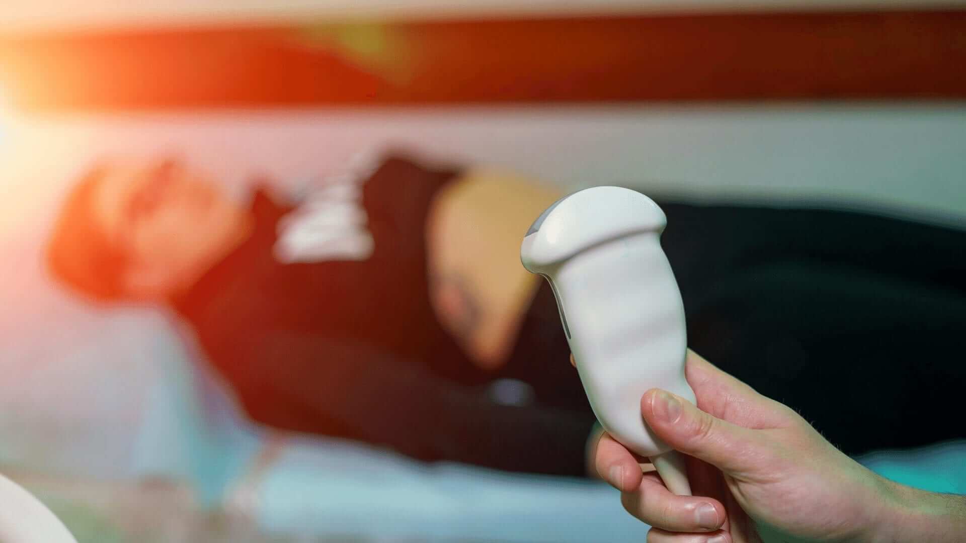
Vascular Ultrasound relies on a handheld portable ultrasound machine and transducer. In the procedure, ultrasonic sound waves are transmitted at high frequencies. By focusing soundwaves at areas of interest, varying echoes return to the scanner, indicating the tissues or liquids encountered. A computer then converts these soundwaves into images, which are recorded and analyzed as informational medical images.
What are Some Typical Uses For Vascular Ultrasound?
Vascular Ultrasound has multiple diagnostic applications:
- Deep Vein Thrombosis = When a blood clot forms in a deep vein. They may develop in the lower leg, thigh, pelvis, or arm.4
- Atherosclerosis = This is a hardening and narrowing of the arteries caused by cholesterol plaques lining the artery over time.5
- Congenital Vascular Malformations = Are abnormally formed blood vessels that one is born with.6
Are There Other Uses For Vascular Ultrasound?
Besides diagnostic capabilities, vascular sonography can determine whether a patient is a favorable candidate for procedures such as angioplasty. The procedure provides information to evaluate the success of surgical procedures such as bypass surgery or graft transplantations. I.e., adequate blood flow to the graft may indicate successful graft transplantation. Additionally, Vascular Sonography procedures may help determine blood flow to tumors and chronic wounds to aid in treatment planning options.
Because these sonographic procedures help identify blood clots and vein blockages, vascular sonography may reveal if blood clots require anticoagulant therapy. This finding is significant because blood clots can travel and embolize to other organs. One well-known danger of blockages in blood flow to the brain is it causes strokes. Therefore, early detection of a blockage can be life-preserving.
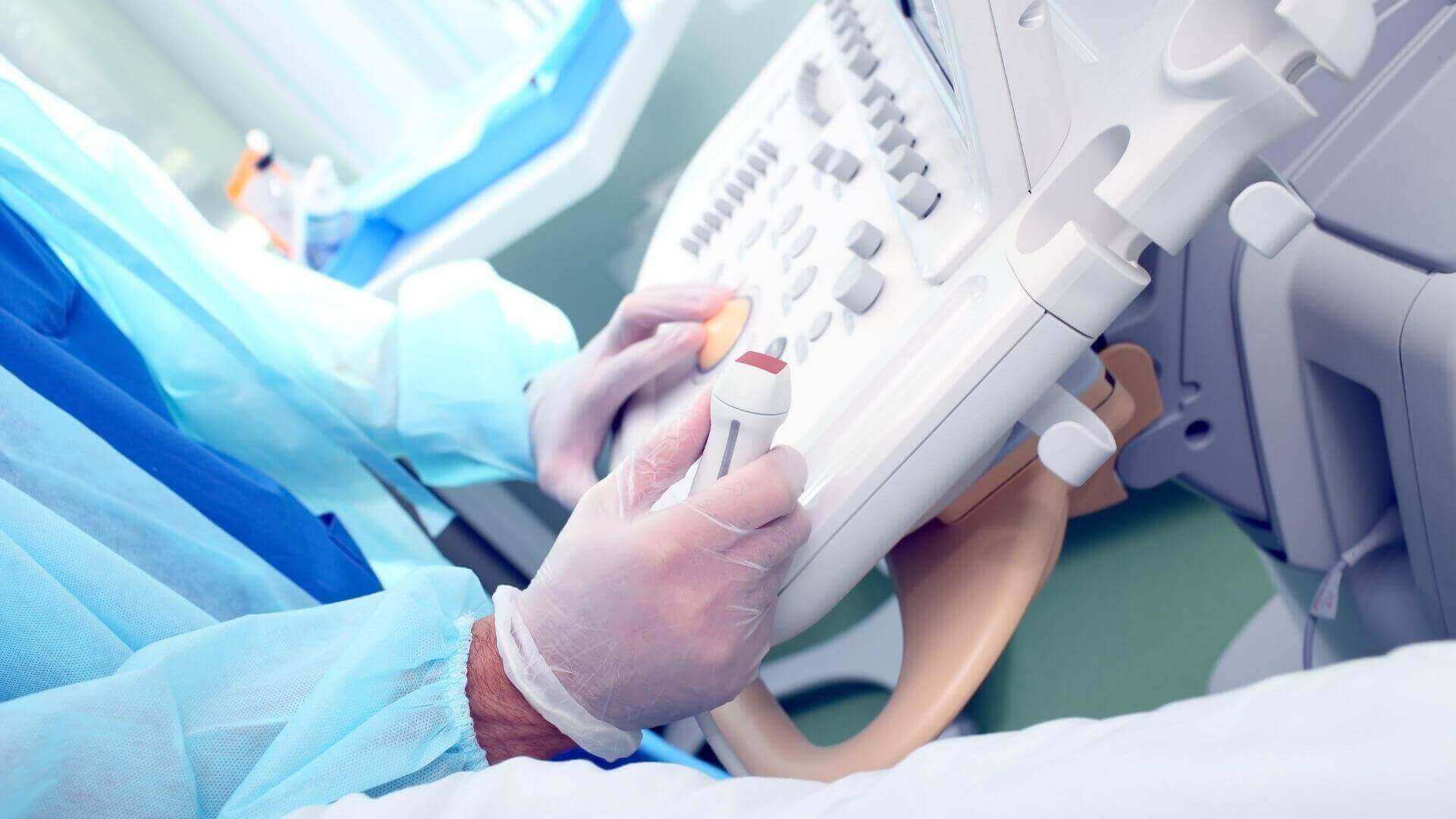
How Are Vascular Ultrasounds Performed?
Generally, Vascular Sonograms are performed in hospital radiology departments or vascular laboratories. However, Vascular Ultrasound units can be portable. Therefore Vascular Sonography can often be carried out at patient bedsides, emergency rooms, or other areas within a hospital or physician’s office.
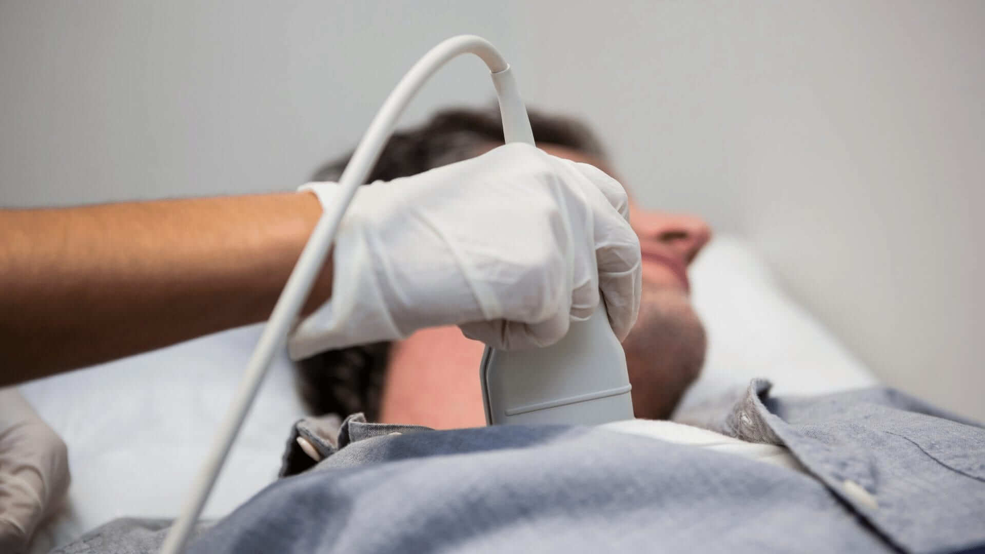
When is This Procedure Recommended?
A primary care physician may recommend this procedure upon detecting abnormal blood flow sounds. Patients with suspected abnormalities in the superficial blood vessels within the arms and legs or those with suspected narrowing of carotid arteries in the neck will benefit significantly from this exam. The exam may also detect the narrowing of the lower abdominal vessels to rule out internal abdominal bleeding following a trauma.
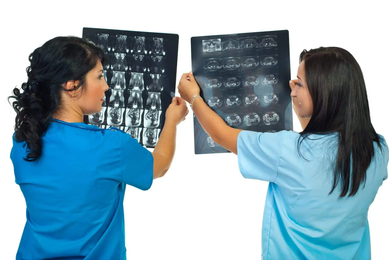
What Are Doppler Images?
Photos acquired through Vascular Sonographic procedures are displayed as gray-scale or Doppler images. These images use varying gray shades to indicate differences in echo strength. Generally, this is shown in the following manner:
- Echoes from the blood of lower vigor appear darker than the surrounding tissues.
- Gray-scale images can depict layers of the vessel walls and can also show real-time arterial motion.
- Expected results generally show normal blood flow in the area under examination.
- Abnormal results generally show abnormalities within the flow of blood.
Summary
The sonogram is used to identify obstructions and abnormalities in blood vessels, including blood clots, arterial plaques, and stenoses. The results might indicate DVT diagnosis, internal bleeding (in the abdomen), or inadequate blood flow to a grafted area.
The next time you think about sound, especially ultrasound, consider what a life-saving procedure sound can be. Do you think a career in ultrasound “sounds” like a “sound” plan? If so, consider contacting Gurnick Academy of Medical Arts today.~
Citations:
1^a, b, c “Understanding Sound—Natural Sounds (U.S. National Park Service).” Www.nps.gov, National Park Service. July 3, 2018. (Accessed August 29, 2022.)
2^a, b —. “Vascular System.” Www.cancer.gov, National Cancer Institute. February 2, 2011. (Accessed September 12, 2022.)
3 Dr. KC. “What Can a Vascular Ultrasound Detect?” Missouri Vein Specialists, Missouri Vein Specialists. June 26, 2020. (Accessed August 29, 2022.)
4 CDC. “What Is Venous Thromboembolism?” CDC Centers for Disease Control and Prevention, U.S. Department of Health and Human Services, 2018. (Accessed September 12, 2022.)
5 WebMD. “What Is Atherosclerosis?” WebMD, WebMD, LLC, November 1, 2021. (Accessed September 12, 2022.)
6 “Congenital Vascular Malformation—Vascular Cures.” Vascular Cures.org, Vascular Cures. (Accessed September 12, 2022.)