The Widespread Diagnostic Medical Benefits of Ultrasound Scans
Date: April 2, 2019
An ultrasound diagnostic imaging technique is performed to view a patient’s internal organs and assess their blood flow through various vessels. An Ultrasound is also beneficial when performed on a pregnant lady so that an obstetrician can check on the developing baby. It does this with high-frequency sound waves, which have no adverse effect on the body.
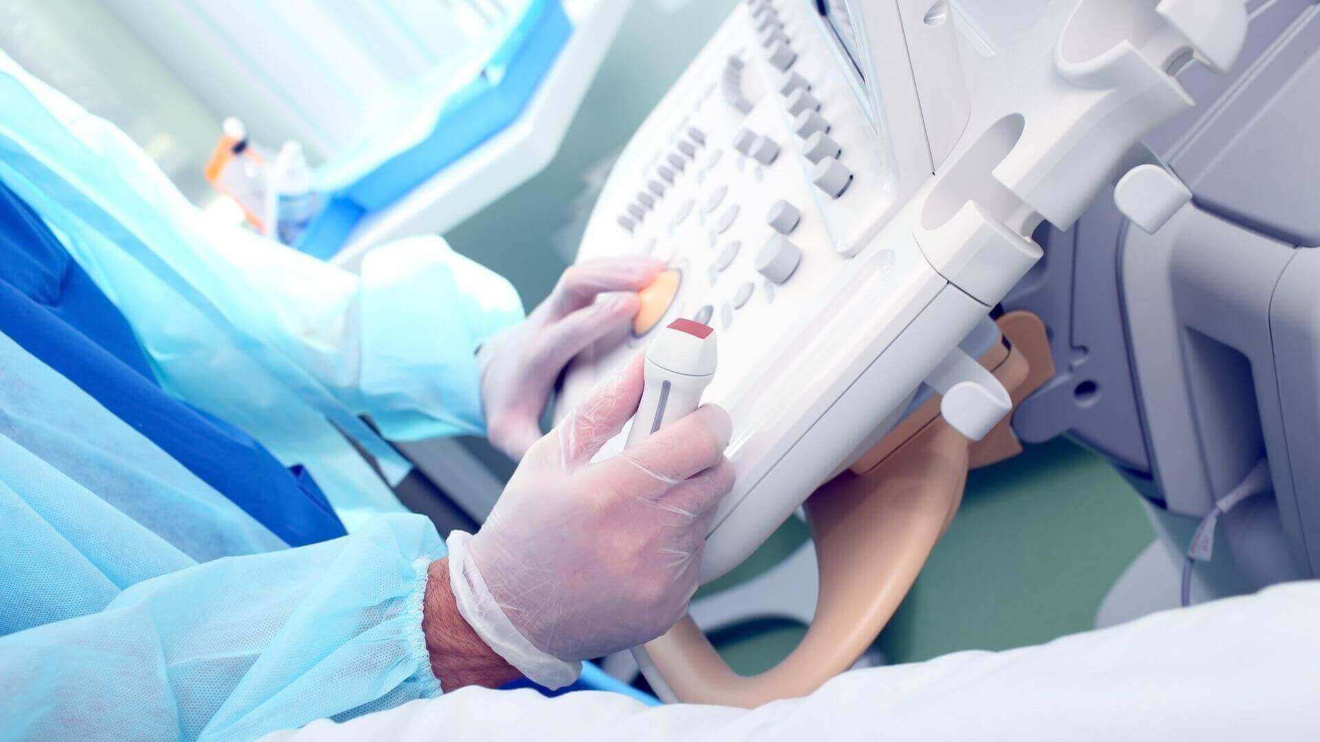
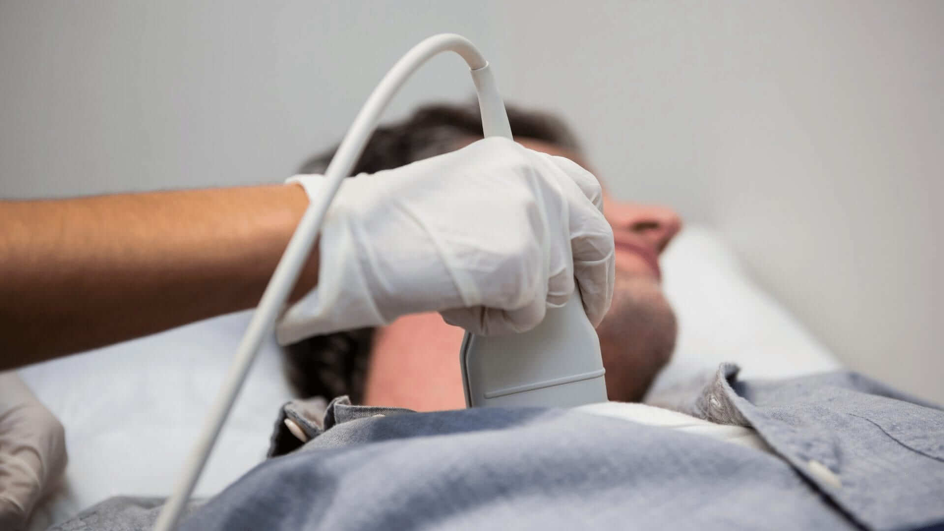
Your average Ultrasound machine or scanner will consist of a computer, a display screen, and a transducer probe used to scan the body. The transducer is the small gadget that the Ultrasound technician will hold in their hand. The transducer is attached to the ultrasound scanner and placed on top of the body area for scanning. The respective area is lubricated with a special gel.
The transducer transmits the sound waves that go through the body, and the internal structure of the body reproduces them as “echoes.” These echoes return to the transducer, which in turn transmits them to the computer part of the Ultrasound scanner, which will exhibit them as real-time visual images.
Ultrasound pictures work on the same basis as the sonar that bats and submarines use to navigate. Since a controlled sound will bounce against any object, its echo perceives the distance the respective body is at, its shape, size, and even its density.
When pressing the Ultrasound scanner’s transducer against the patient’s skin, it starts directing a stream of muted and high-frequency sound waves into the body. The sound waves echo from the body’s fluids, tissues, and organs and are picked up by the transducer, which records the tiny changes in the sound’s pitch and direction. These echoes are instantly measured and displayed by the computer, generating a real-time picture on the technician and doctors’ display screen.

Ultrasound images have proven to be a suitable method of guiding minimally invasive procedures and a handy diagnostic instrument in Obstetrics. Ultrasound scanner images examine the body to detect muscles, tendons, ligaments, joints, and soft tissue problems. Ultrasound images will often demonstrate movement, function, and anatomy and allow radiologists to analyze an assortment of conditions and assess damage after an injury or illness.
Ultrasound scanning is also a suitable method of examining the body’s internal organs, including the heart, liver, gallbladder, spleen, pancreas, kidneys, and bladder. Ultrasound scans help determine the sources of pain, swelling, or infection a patient may suffer. Since the Ultrasound images are captured in real-time, they demonstrate the movement of internal tissues and organs and allow physicians to observe the patient’s blood flow and heart valve functions.
This versatility of the Ultrasound scanner helps to diagnose the damage after a heart attack or other illness. Gallstones and kidney stones are easily spotted. Ultrasound scans will identify fluid, cysts, tumors, abscesses in the abdomen or liver and detect impaired blood flow from clots or arteriosclerosis in the legs. Ultrasound images also evaluate superficial structures such as the thyroid gland and scrotum. Most Ultrasound examinations take forty-five minutes or less.