
MRI vs. Ultrasound: Two Vital Branches of Radiology
Date: September 29, 2023
Radiology involves the study of internal organ images within the human body. According to the Oxford Languages Dictionary, it is the science dealing with X-rays and other high-energy radiation, especially for diagnosing and treating disease.1
Before the advent of diagnostic imaging, only a patient’s death allowed doctors to study the body’s internal organs and structures comprehensively. However, Wilhelm Röntgen’s accidental discovery of the X-ray beam in 1895,2* gave healthcare providers “front row seats” to view inside the human body while a patient was still living. What’s more, radiologic imaging allows physicians to treat patients and ward off early fatalities.
Growth of Imaging
In the beginning, initial diagnostic procedures were crude. Mainly, imaging consisted of flat sheets of photographic film to provide images to practitioners. However, modern-day radiology is no longer confined to simple images. By contrast, healthcare providers and patients get views once only dreamed of. Radiologists can glimpse at heart tissue, look at the brain, and view inside the blood vessels.
Additionally, today’s radiology encompasses many different types of diagnostic imaging methods. Radiologic professionals may employ diagnostic and interventional radiology to examine the body.3

Diagnostic Radiology VS Interventional Radiology
In sweeping terms, diagnostic radiology includes procedures such as computed tomography (CT), fluoroscopy, magnetic resonance imaging (MRI), and nuclear medicine. It further comprises X-rays, positron emission tomography (PET scans), and ultrasound (UT).3 While interventional radiology encompasses such procedures as angiography, embolization, tumor embolization, tumor ablation, vertebroplasty, needle biopsies of different organs, uterine artery embolization, and venous access catheter placement. These include ports and PICCs.3 Although interventional radiology is essential, this article focuses on two specific diagnostic radiology procedures.
Diagnostic Radiologic Procedures Examined
Specifically, we’ll examine two diagnostic radiologic procedures: magnetic resonance imaging (MRI) and ultrasound (UT).3 Unlike other radiologic techniques such as the X-ray, MRI, and ultrasound, imaging methods don’t require ionizing radiation. For this reason, they are considered relatively safe. Therefore, such techniques are fast becoming popular–even preferred medical procedures.
ONE: Magnetic Resonance Imaging (MRI)

First, let’s look at Magnetic Resonance Imaging. In brief, the MRI is a non-invasive diagnostic technique that combines a large, powerful magnetic field with radio frequencies. This emission-based imagining produces detailed pictures of organs, soft tissues, bones, and other internal body structures. Moreover, the MRI procedure allows physicians to evaluate and view diseased areas that may not be assessed adequately by other imaging methods.
Additionally, the MRI scan produces the highest quality soft-tissue contrasts of all other forms of diagnostic imaging. For this reason, such scans are especially beneficial for imaging the brain, spine, and musculoskeletal system.
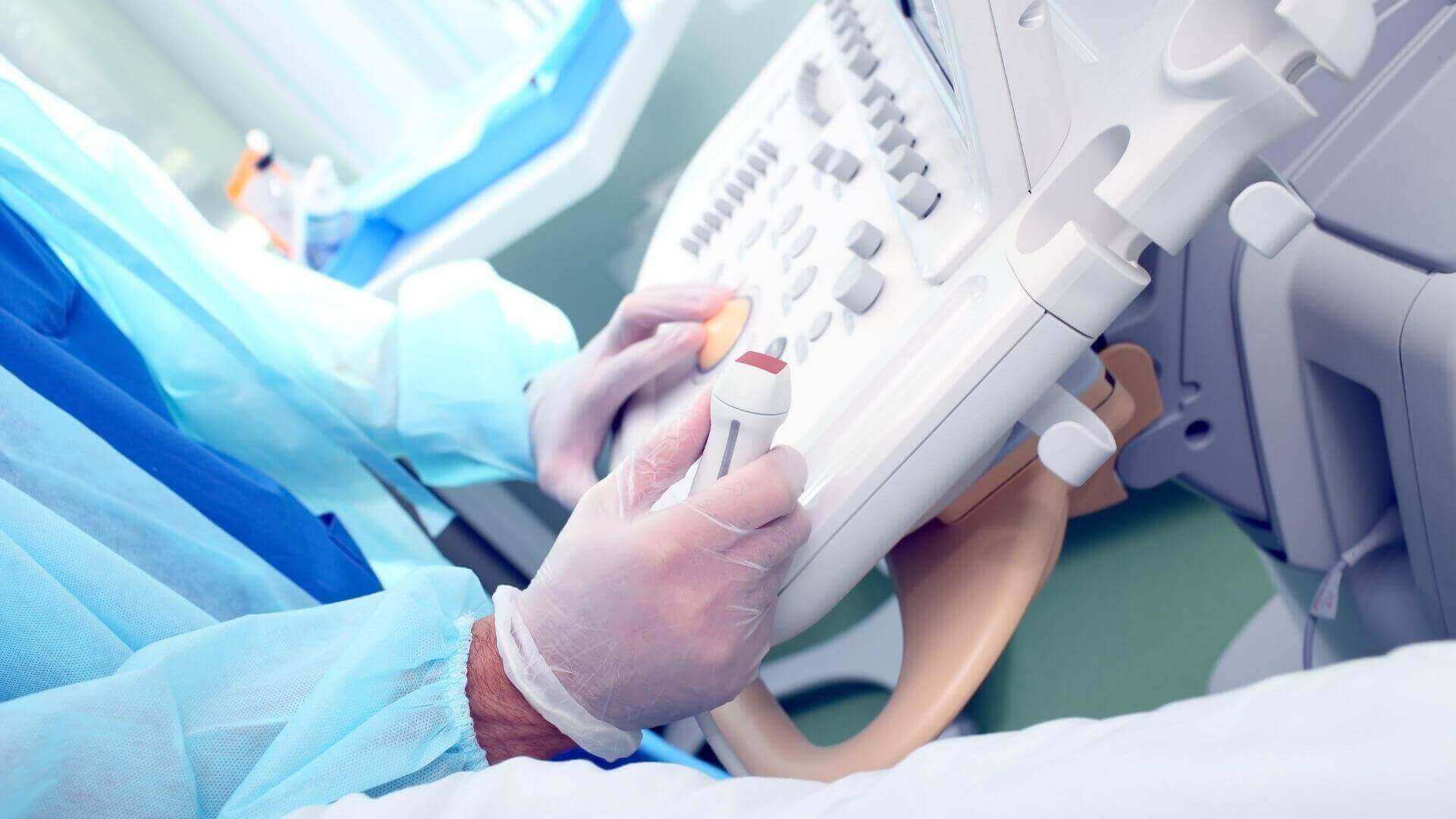
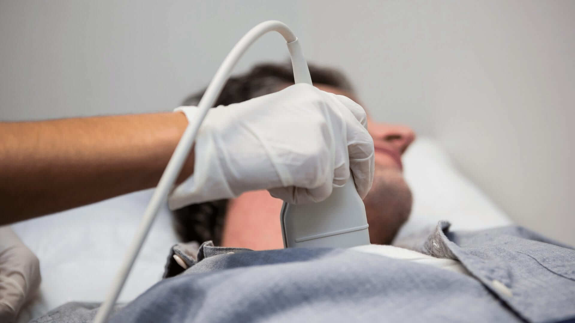
TWO: Ultrasound (UT)

Second, ultrasonography, or ultrasound technology, is a useful diagnostic modality employing high-frequency sound waves to visualize various organs and organ systems. In short, the ultrasound procedure is an example of reflection imaging. Moreover, ultrasounds are commonly used to examine the abdomen, small parts, veins, arteries, and male and female reproductive systems.
By and large, ultrasound imaging plays a critical role in obstetrics and fetal imaging. Ultrasound is used to rapidly diagnose medical, surgical, obstetrical, and gynecological problems. However, ultrasound has many more uses than just the ones explained above.
MRI and UT Together
MRI and ultrasound imaging modalities occupy a vital position in radiology. Not only do the techniques produce computerized images, but they also help diagnose patient ailments. Moreover, both have narrowed the bridge between undetected illnesses. Subsequently, they have helped healthcare providers establish care plans at the onslaught of disease, injury, or illnesses.
Employment of MRI and UT Technologists
As the field expands, so does employment. According to the Bureau of Labor and Statistics (BLS), overall employment for MRI Technologists is projected to grow upward of nine percent from 2020 to 2030. This increase is about as fast as the average for all occupations. Consequently, approximately 20,800 openings for MRI Technologists are projected yearly over the next decade.4 It’s safe to say the demand for Diagnostic Imaging Technologists is significantly increasing. Moreover, workforce shortages in the industry attract a wide range of individuals as they launch radiologic field vocations.4
Likewise, the BLS cites the overall employment of medical sonographers as projected to grow ten percent from 2022 to 2032,5 much faster than the average for all occupations. Moreover, there are approximately 9,600 openings5 for diagnostic medical sonographers each year, on average, over the next decade. Many of these openings are expected to result from the need to replace workers who transfer to different occupations. Others are because many workers are exiting the labor force, such as retiring.5
Are Either of These Careers For You?
Do you think either an MRI or UT career path is for you? Gurnick Academy offers 16 unique programs across our various campuses. So, whether you’re looking for continuing education courses (CECs), certificates, diplomas, or degrees, Gurnick Academy has the perfect diagnostic imaging program to help you reach your educational goals. Explore our multiple campus locations and individual program pages to find the right fit for you. Talk to one of our experts today. ~
*Röntgen learned that the mysterious light could pass through almost all solid objects through experimentation. Because he did not know what the rays were, he called them X-rays, where the x means “unknown.”2
Citations
1 “Radiology.” Oxford Languages, Oxford University Press. (Accessed June 9, 2023.)
2^a, b Daley, Bonitto. “History of Radiology.” Radiology Affiliates Imaging, Apr. 2021. (Accessed June 9, 2023.)
3^a, b, c, d “Imaging and Radiology: MedlinePlus Medical Encyclopedia.” Medlineplus.gov, U.S. Department of Health and Human Services National Institutes of Health. (Accessed June 9, 2023.)
4^a, b Bureau of Labor Statistics, U.S. Department of Labor, Occupational Outlook Handbook, Radiologic, and MRI Technologists. (Accessed March 18, 2022.)
5^a, b, c Bureau of Labor Statistics, U.S. Department of Labor, Occupational Outlook Handbook, Diagnostic Medical Sonographers and Cardiovascular Technologists and Technicians. (Accessed September 12, 2023.)