Ultrasound Technology
Date: October 18, 2019
Ultrasound Technology is a high-energy sound wave that frequency is higher than the upper limit of humans’ hearing capacity. It lies somewhere around 20,000 kilohertz. Ultrasound scanning is the most sought-after form of scanning in medicinal science today, thanks to its ability to produce high-quality images that help physicians diagnose the patient’s body condition.
In medicinal terms, the procedure is called ultrasonography. It is done by passing high-frequency sound waves through the patient’s body to get visual images of the internal organs, tissues, and blood flow. These images help physicians analyze the patient’s body condition and develop an idea of the abnormality causing the problem. This procedure is commonly called an ultrasound scan.
An ultrasound scan is done by passing through sound waves that have a frequency higher than 20,000 kilohertz. It ranges from 3 to 15 megahertz, as high frequencies produce high-resolution images. Superficial organs such as muscles, tendons, breasts, and genitals don’t require significant penetration by the sound waves and are scanned in very high frequency (7-15 megahertz) to get high-resolution images.
Deeper organs such as the liver, kidney, and pancreas cannot be reached with very high-frequency waves and are imaged using a lower frequency (1-6 megahertz) to get low-resolution images with greater penetration. So, it’s the sonographers’ choice at the end of the day to choose which frequency to get the scan done, depending on the physician’s need.
Ultrasound Technology is used in various branches of medicine. This includes cardiology, endocrinology, gastroenterology, gynecology, obstetrics, urology, musculoskeletal, and ophthalmology. Specialty images can be produced in the case of different branches of medicine by using a specialty transducer. An essential and most common usage of ultrasound scans is to check the fetus’s development during pregnancy.
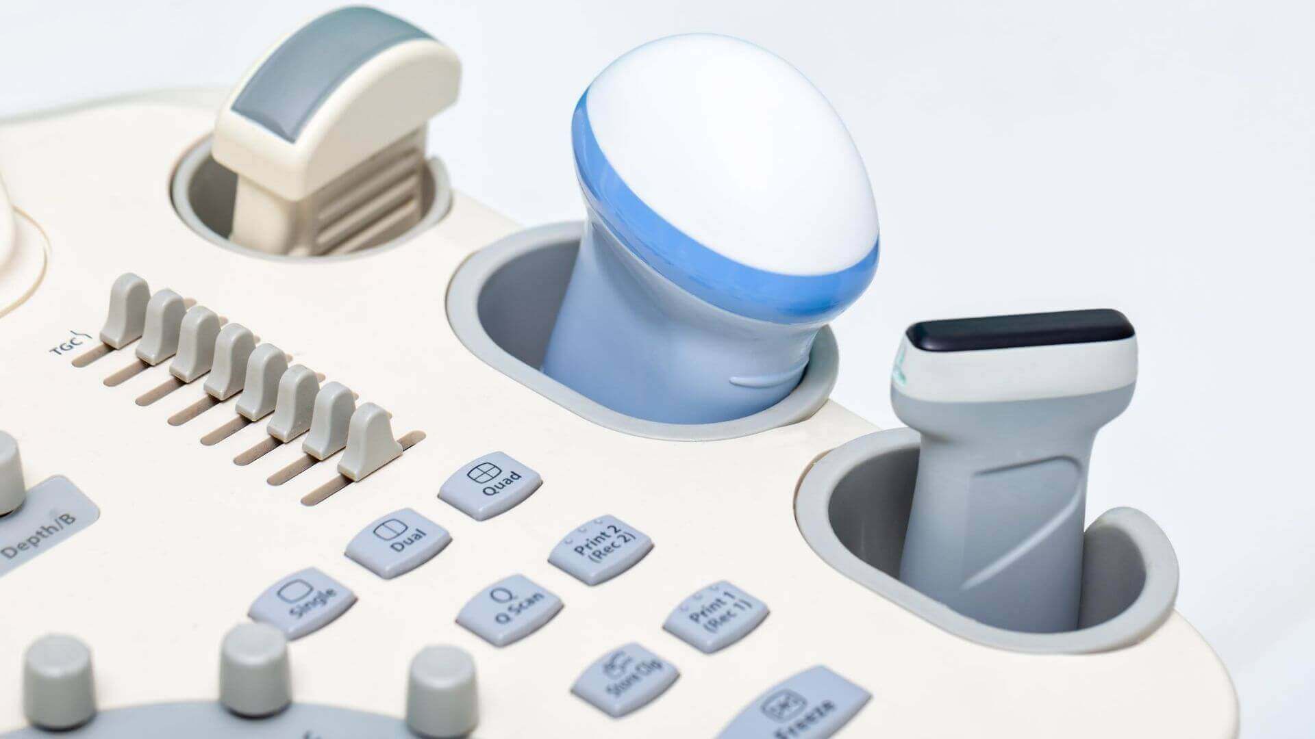
An ultrasound scan should be done only by qualified sonographers who have professional training. It requires a more in-depth understanding of anatomy, physiology, equipment usage, instrumentation techniques, and patient care. The sonographer must also arrive at decisions during the scan regarding the frequency used and other related things. An ultrasound scan usually has three stages. Production of the sound wave, reception of the echo, and interpretation of the echo received.
A qualified sonographer should be able to interpret the echoes received and should be able to explain the physician. Apart from the usual diagnostic purposes, ultrasound can also be used for various other purposes. It can be used in the field of dentistry to clean teeth.
It can generate heat in the body’s softer tissues during occupational therapy, physical therapy, and in treating some forms of cancer. Focused ultrasound may also be used to treat both benign and malignant tumors. It can also be used to break kidney stones or renal calculi.